The Charles T. Campbell Eye Microbiology Lab
UPMCUniversity of Pittsburgh Schools of the Health Sciences
HomeContact InformationLab Diagnostic TestingAntibiotic SusceptibilityAntimicrobial TherapyCurrent ResearchPhotos


Lab Diagnostic Testing: Chlamydia

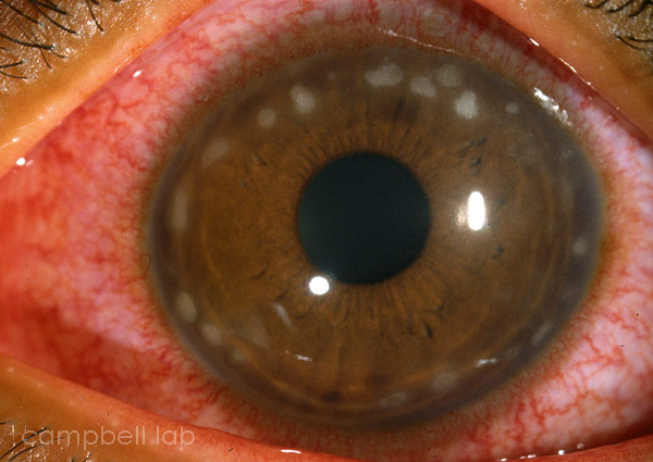
Conjunctivitis due to Chlamydia can be an acute or chronic disease. Chlamydia trachomatis is an intracellular parasite that can be diagnosed in the laboratory with 1) smears (giemsa, immunofluorescence), 2) cell culture, 3) serology, 4) polymerase chain reaction (PCR) for DNA, and 5) Nucleic Acid Amplification Testing (NAAT) for rRNA (Gen-Probe® Aptima®). Although our laboratory has utilized all these tests at some time, we currently test for Chlamydia with the giemsa stain and NAAT testing. Other laboratories may favor other tests, but we suggest that ophthalmologists only choose tests that are 100 percent specific (avoid positive tests that are actually negative), and highly sensitive (positive results are truly positive). Testing with cell culture is not necessary if PCR or NAAT testing is available.

Specimen Collection
Smears (Giemsa)

PCR or NAAT

Specimen Collection
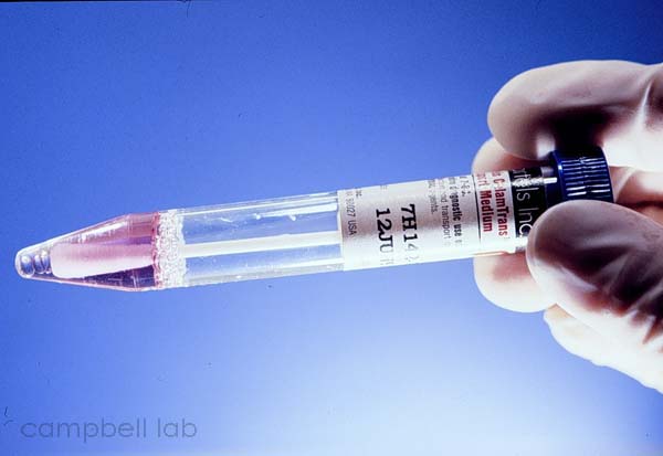
Specimens are directly collected by vigorously swiping the exposed conjunctiva with a plastic soft-tipped applicator. Cornea samples are not necessary. Topical anesthetic can be applied to the conjunctiva but this is optional. Collected samples are placed in 2.0 ml of Chlamydial transport medium. We have had great success with Bartels ChlamTrans™ Chlamydial transport medium and recommend its use. DO NOT USE VIRAL TRANSPORT MEDIUM THAT MAY CONTAIN ANTIBIOTICS THAT INHIBITS CHLAMYDIA GROWTH IN CELL CULTURE. Chlamydia is very fastidious and will not survive unless refrigerated (short-term) or frozen (long-term) (-75 degrees C). All samples for cell culture must be transported immediately to the laboratory. PCR and NAAT can be processed from the transport medium.

Smear specimens should be obtained with a conjunctival spatula. NEVER USE A SWAB (soft-tipped applicator) TO OBTAIN A SMEAR FOR CYTOLOGY. After applying topical anesthetic, specimens are directly collected by firmly scraping the exposed conjunctiva. These specimens are transferred to glass microscope slides, air-dried, and allowed to remain at room temperature.

Transport medium
(Click on image to enlarge)
Transport medium

Smears (Giemsa)
Giemsa – The giemsa stain is used to examine conjunctival specimens for cytology and microorganisms. It could provide pertinent information in about one hour. After a specimen is obtained, the slides are air-dried, fixed with methanol, and placed in giemsa stain. Under microscopy, a conjunctival specimen from a patient with ocular Chlamydia would contain many epithelial cells, many polymorphonuclear cells, moderate mononuclear cells, and a few plasma cells. This cytology alone could support a chlamydial diagnosis. Chlamydial inclusion bodies (initial, elementary) can also be observed in the cytoplasm of the epithelial cell especially in newborns 5 to 20 days after birth but less frequently in young and older adults. Although the giemsa stain is a powerful tool, it rarely is offered because of the lack of experienced personnel.

Chlamydia inclusion bodies
(Click on image to enlarge)
Chlamydia inclusion bodies

PCR or NAAT
We highly recommend PCR or NAAT testing for the detection of chlamydial DNA from ocular specimens. The problems are less with transportation of these specimens because live organisms are not necessary. Still, samples should be refrigerated and transported to the laboratory as soon as possible to avoid any possible enzymes that may degrade any DNA or rRNA. We advise ophthalmologists to make the effort to locate PCR or NAAT testing.

Samples are prepared for NAAT processing with the following proceudure: Vigorously vortex the sample collected in Chlamydia transport medium. Place 1.0 ml of sample into a 2.0 ml polypropylene micro tubes with a conical bottom. Centrifuged at 13,000 rpm in a refrigerated centrifuge at 4oC. Using the Gen-Probe® Aptima® NAAT Kit Unisex collection blue swab, a specimen is collected from the conical apex where a pellet may have formed from centrifugation. The Unisex collection swab is placed in a tube of Gen-Probe® Aptima® swab transport medium and submitted for NAAT testing.

Top of Page


Chlamydia Keratoconjunctivitis
(Click on image to enlarge)
Chlamydia Keratoconjunctivitis