|

Lab
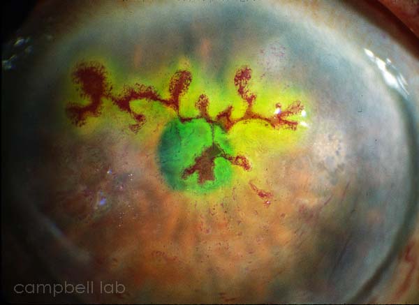
Diagnostic Testing: Herpes Simplex Virus
The presence
of herpes simplex (HSV) from ocular specimens is essential for prompt
and accurate therapy. HSV can be detected from ocular samples with
cell
culture and ELVIS
(Enzyme Linked Viral Induced System).
Specimen
Collection
Cell
Culture
ELVIS
Specimen
Collection
Specimens are directly collected by vigorously swiping the exposed
conjunctiva with a plastic soft-tipped applicator. Cornea samples
using soft-tipped applicators and spatulas can also be obtained
to maximize the yield of viable HSV and antigen. Topical anesthetic
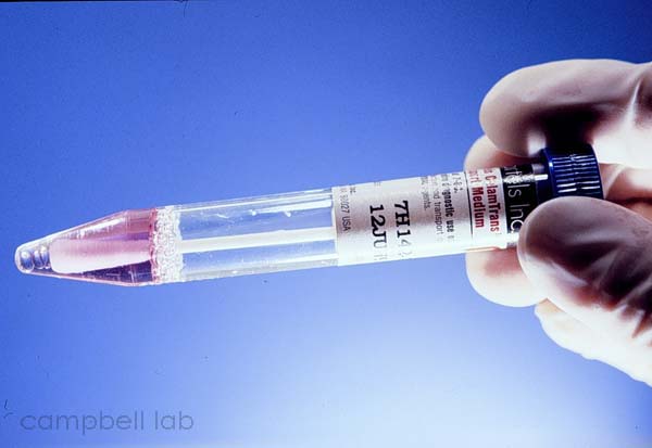
can be applied to the conjunctiva and should be applied to the cornea.
Collected samples are placed in 2.0 ml of viral transport medium.
We have had great success with Bartels Chlamydial transport medium
and recommend its use. Viral culturettes can also be used for transportation
to the laboratory and these can be transferred to the viral medium.
All laboratory testing can be processed from the 2.0 ml of chlamydial
transport medium. Herpes simplex virus can be fastidious and should
be transported to the laboratory without unnecessary delay.

Cell
Culture
The "gold" standard for HSV laboratory testing is cell
culture. Collected samples are layered on a monolayer of cells in
test tubes. If present, HSV will present as cytopathic effect of
rounded cells. The cytopathic effect of can be confirmed for the
presence of antigen by EIA (i.e. Herpchek). We use A549 cells as
the cell-line for testing HSV. These are a human carcinoma continuous
cell-line. When samples are positive for HSV, cytopathic effect
is generally present within one to three days. It is rare, but sometimes
one to two weeks is necessary to isolate HSV in cell culture. Cell
culture will confirm an HSV diagnosis but it may not provide timely
results for immediate patient care. All virology laboratories can
offer cell culture isolation for HSV.

CPE observed in cell culture (A549 cells).

ELVIS
ELVIS (Enzyme Linked Viral Induced System) is another cell
culture test but the results are ready in ONE day. Cells infected
with HSV commence a cascade of reactions that results in the accumulation
of beta-galactosidase in the cells. Viral samples are layered on
the specially engineered cell line in a shell vial and centrifuged.
The vial is incubated for one day (overnight), fixed, and stained
with a substrate that reactions with the beta-galactosidase. The
reactions result in a blue-color change in the cells that are observed
with an inverted microscope. ELVIS highly correlates with Herpchek
but negative results also must be confirmed with standard cell culture.
ELVIS testing may also be limited in its availability. We highly
recommend ELVIS testing for diagnosing ocular HSV infection.

Positive
stained ELVIS cells.

|